CT Brain Imaging: Techniques and Clinical Insights
Intro
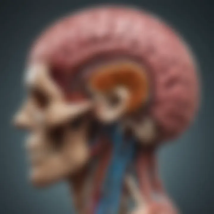
CT brain imaging has transformed the landscape of neurosciences and clinical diagnostics. Its ability to produce high-resolution images quickly provides invaluable information for diagnosing a range of neurological disorders. This article aims to explore not just the technology behind CT scans, but also their clinical applications and the interpretation of the images they generate.
In recent years, advancements in imaging techniques have improved the accuracy and efficiency of CT scans. Understanding these techniques is essential for researchers and practitioners alike. This narrative outlines the current state of CT brain imaging, its methodologies, and its role within the broader field of neuroimaging.
Preamble to CT Brain Imaging
CT brain imaging plays a critical role in modern medicine, particularly in the field of neurology. By utilizing computed tomography, healthcare providers can obtain detailed cross-sectional images of the brain. This imaging technique is vital for diagnosing a variety of neurological conditions.
One important element of CT brain imaging is its speed. The ability to quickly obtain images allows clinicians to make timely decisions, especially in emergency situations. For instance, in cases of trauma or suspected stroke, rapid imaging can significantly influence patient outcomes. The efficiency of CT scans in these scenarios cannot be overstated.
Furthermore, CT brain imaging provides great detail when evaluating bony structures and acute injuries. The precision of the images assists neurologists in spotting fractures, hemorrhages, and other critical issues. This exceptional capacity to visualize anatomical details enhances diagnostic accuracy.
While the advancements in CT technology are noteworthy, it is also important to discuss considerations such as radiation exposure. As these scans involve the use of ionizing radiation, understanding the risk versus benefit is crucial when deciding to perform a CT scan. Appropriate protocols must be in place to minimize unnecessary exposure while ensuring diagnostic quality.
In summary, the introduction of CT brain imaging is foundational to understanding its clinical applications. As the following sections will elucidate, there are numerous advantages and specific uses of this technology, making it an invaluable tool in the diagnosis and management of neurological conditions.
"The advent of CT imaging was revolutionary, providing unparalleled insights into the human brain."
The Physics of Computed Tomography
The physics behind computed tomography (CT) is foundational for understanding how this imaging technique works. This section explains the principles that govern CT technology, the mechanics of x-ray production, and the methods used to reconstruct data into images. Knowledge in these areas is crucial for professionals in the medical imaging field.
CT technology relies on principles from physics to create detailed images of the human body, particularly the brain. By understanding these principles, one can appreciate the advantages CT imaging offers in clinical settings, especially in emergency scenarios where time is critical.
A crucial aspect of CT technology is that it involves understanding how x-rays are generated and how they interact with tissues of varied densities. This enables the differentiation between various structures within the body and is a key reason for its widespread use in detecting neurological conditions.
Principles of CT Technology
CT technology utilizes a rotating x-ray machine and multiple detectors to capture images from different angles. Each rotation generates a series of x-ray images, which are processed to create cross-sectional images of the brain. The science behind CT scanning involves the conversion of x-ray attenuation into digital data.
When x-rays pass through the head, they are absorbed differently by various tissues. Dense materials, like bone, absorb more x-rays compared to softer tissues, such as cerebral tissue. This differential absorption creates a contrast that is key to imaging. The data collected by the detectors is then processed through algorithms to reconstruct the images.
X-Ray Production and Detection
The process begins with the generation of x-rays from a tube. X-ray production occurs when high-energy electrons collide with a target material, usually tungsten. This collision results in the emission of x-rays. Once generated, these x-rays are directed towards the patient.
It is important to understand that different tissues absorb x-rays at different rates. The resulting pattern of absorption information is collected by detectors positioned around the patient's head. The data is subsequently converted into electrical signals for further processing.
The effectiveness of x-ray detection depends on the design of detectors. Modern detectors are able to capture multiple high-quality images in a short time, which enhances the diagnostic process. This rapid image acquisition is essential in emergencies, as it reduces the time needed to obtain critical information.
Data Reconstruction Algorithms
Once the data has been collected, it undergoes reconstruction through sophisticated algorithms. These algorithms transform the raw data into coherent images. The most common approach is the filtered back-projection algorithm, although advanced techniques like iterative reconstruction are gaining popularity due to their superior image quality.
One advantage of newer reconstruction methods is improved noise reduction. This is particularly beneficial in low-dose scans, where reducing radiation exposure is vital. However, it's essential to strike a balance between image quality and radiation dose, as excessive radiation can pose risks to patients.
The integration of advanced data reconstruction algorithms has revolutionized the way CT images are produced, enhancing resolution and detail, which is crucial for accurate diagnosis.
In summary, the physics of computed tomography plays a vital role in the quality and effectiveness of brain imaging. By understanding the principles of CT technology, the production and detection of x-rays, and the data reconstruction processes, professionals can make informed decisions that significantly affect patient outcomes in clinical practice.
Types of CT Scans in Neurology
CT scans serve as an essential tool in neurology, allowing for detailed visualization of brain structures and pathologies. Understanding the various types of CT scans is crucial for clinicians to effectively diagnose and manage neurological conditions. Each scan type provides unique advantages and limitations, guiding the choice of technique based on the clinical scenario.
Non-Contrast CT Scans
Non-contrast CT scans are the most basic form of CT imaging used in neurology. Their primary advantage is speed, allowing quick assessments of acute conditions. These scans are particularly beneficial in emergency settings, aiding in the rapid detection of hemorrhages, fractures, and other urgent issues. They work by producing images without the use of contrast agents, which minimizes potential adverse reactions in patients.
Key points about non-contrast scans include:
- Speed: Allows immediate imaging which is crucial in acute settings, especially for trauma cases.
- Immediacy: Can be completed quickly, providing prompt results.
- Cost-effective: Generally less expensive than contrast-enhanced scans.
Limitations exist, such as the inability to visualize subtle changes in soft tissues and vascular structures. This type of CT is excellent for assessing bone injury but may not reveal everything needed for a comprehensive diagnosis.
Contrast-Enhanced CT Imaging
Contrast-enhanced CT imaging involves the injection of a contrast medium, usually iodine-based, to improve the visibility of certain structures within the brain. This technique highlights vascular structures and enhances the differentiation of various tissues. It is particularly useful in evaluating tumors, abscesses, and vascular malformations.
Some benefits of contrast-enhanced scans include:
- Enhanced Visualization: Differentiates between tumors and normal tissues, aiding diagnosis and treatment planning.
- Vascular Imaging: Helps assess blood vessels and detect conditions like aneurysms and stroke.
- Guidance for Procedures: Improves accuracy when performing interventions, such as biopsies.
However, contrast-enhanced CT scans carry some risks, including allergic reactions to the contrast material and potential impacts on patients with compromised kidney function. Thus, careful consideration is always needed prior to administration.
CT Angiography
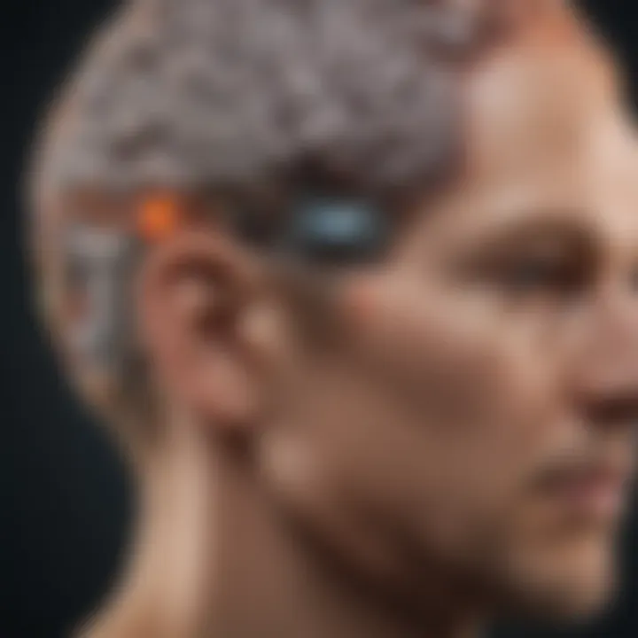
CT angiography is a specialized form of contrast-enhanced CT scan that focuses on visualizing blood vessels. It combines standard CT imaging with contrast material to create detailed images of the blood vessels in the brain. This technique plays a vital role in diagnosing vascular conditions such as aneurysms, dissections, and stenosis.
Important points about CT angiography include:
- Detailed Vascular Imaging: Provides superior visualization of blood vessels, crucial for diagnosing vascular diseases.
- Non-Invasive: Unlike traditional angiography, it does not require more invasive catheter insertion, making it safer for patients.
- Rapid Acquisition: Offers quick imaging, a necessity in acute stroke cases or when assessing vascular emergencies.
Despite its benefits, the reliance on iodine-based contrast can still pose risks. It is essential to evaluate patient history and renal function before performing CT angiography.
In summary, each type of CT scan has unique features that cater to specific clinical needs in neurology. The selection is often a balancing act between speed, detail, safety, and patient history. Adequately understanding these variations aids clinicians in making informed decisions that ultimately improve patient outcomes.
Clinical Indications for CT Brain Imaging
The role of computed tomography (CT) brain imaging in clinical practice cannot be overstated. Its applications are diverse, influencing both diagnosis and treatment options for various neurological conditions. This section focuses on three primary clinical indications: trauma assessment, stroke diagnosis, and tumor evaluation. Understanding these applications helps clarify the importance of CT scans in modern medicine.
Trauma Assessment
Trauma assessment through CT imaging is critical in emergency medicine. When patients present with head injuries, a swift diagnosis is essential. CT scans provide rapid, high-resolution images of the brain. This capability is instrumental in identifying fractures, hemorrhages, and contusions. The speed of CT scans allows medical teams to make informed decisions quickly, potentially saving lives.
For example, in cases of severe head trauma, a CT scan can identify subdural hematomas or intracranial bleeding. Prompt identification of these conditions often dictates the urgency of surgical intervention. The American College of Radiology recommends CT as the primary imaging modality for initial trauma assessment.
Stroke Diagnosis
In the context of stroke, time is of the utmost essence. CT imaging plays a pivotal role in differentiating between an ischemic stroke and a hemorrhagic stroke. A non-contrast CT scan is typically the first imaging modality used. It can quickly reveal the presence of blood in the brain, indicating a hemorrhagic event.
On the other hand, if ischemic stroke is suspected, advanced CT techniques such as CT angiography may be employed. This aids physicians in assessing blood vessel patency and can inform treatment decisions regarding thrombolysis or thrombectomy. Patients diagnosed with stroke can often benefit from rapid CT imaging, enhancing the chances of better clinical outcomes.
Tumor Evaluation
CT brain scans are also indispensable in the evaluation of brain tumors. They provide detailed information regarding the size, location, and extent of brain lesions. For newly diagnosed tumors or recurrent disease, CT imaging facilitates the assessment of treatment effectiveness.
Specifically, a contrast-enhanced CT can help delineate tumor boundaries and identify associated edema. Recognizing these factors is vital for devising an appropriate treatment plan. Furthermore, follow-up scans can track changes over time, providing valuable insights into the tumor's response to therapy.
"Early detection and effective imaging can be life-saving, especially for conditions like stroke or brain tumors where timing is critical to treatment success."
In summary, the clinical indications for CT brain imaging are central to effective diagnosis and management of neurological emergencies. These applications illustrate not only the technical advantages of CT scans but also their necessity in ensuring timely, precise medical interventions.
Advantages of CT Brain Imaging
CT brain imaging offers several significant advantages, making it an invaluable tool in modern neurology. Within the context of this article, understanding these advantages aids in appreciating the role CT scans play in clinical settings. The benefits include speed and accessibility, cost-effectiveness, and the capability to detail bone and dense structures. These aspects contribute to better patient care and improved diagnostic accuracy, making CT an essential component of neuroimaging.
Speed and Accessibility
CT scans provide rapid imaging solutions in emergency situations. The technology enables quick acquisition of images, which is crucial when assessing conditions such as strokes or trauma cases. Typical scan times can be mere minutes, allowing for efficient evaluation and timely intervention. This speed not only benefits the patients but also enhances hospital workflow.
Moreover, CT scanners are widely available in many healthcare facilities, from urban hospitals to rural clinics. This accessibility ensures that patients can receive accurate imaging when they need it most, regardless of their location.
Cost-Effectiveness
In the realm of healthcare, cost-effective solutions are critical. CT imaging is generally more affordable than other imaging modalities, such as MRI. The lower capital costs associated with CT machines and the quicker scan times contribute to its economical advantages. Insurance coverage for CT scans is often more favorable, which can ease the financial burden on patients.
This cost-effectiveness does not come at the expense of quality. On the contrary, CT provides excellent diagnostic information in many cases, making it a practical choice in both acute settings and routine evaluations. Patients benefit from the timely decision-making that affordable imaging facilitates, contributing to overall better health outcomes.
Detailing Bone and Dense Structures
CT imaging is particularly adept at visualizing bone and dense structures, a significant advantage in various medical situations. The high-resolution images produced by CT scans allow for accurate assessment of fractures, tumors, and other conditions that involve hard tissues.
For example, in trauma cases, clinicians can quickly identify skull fractures and intracranial hemorrhages, which are essential for determining the appropriate course of treatment. The ability to differentiate these injuries aids in minimizing the risk of complications.
CT technology continues to evolve, enhancing capabilities to provide even more detailed images, thereby improving clinical outcomes across a spectrum of neurological conditions.
CT imaging is often the first choice in emergency assessments due to its rapidity and high-quality representations of cranial anatomy.
These advantages solidify CT brain imaging as a cornerstone in neurology, offering prompt and comprehensive insights that are critical for effective diagnosis and management.
Limitations of CT Imaging in Neurology
CT imaging is a widely used technique in neurology, but it is important to acknowledge its limitations. Understanding these constraints is essential for medical professionals who utilize CT scans for diagnosis and treatment strategies. Identifying the drawbacks not only helps to manage patients better but also guides ongoing research and technological improvements in imaging modalities.
Radiation Exposure Concerns
One significant limitation of CT imaging is the radiation exposure that patients may incur during scans. CT scans utilize ionizing radiation to create detailed images. Although the actual risk of radiation-induced health problems appears to be low for most patients, especially relative to the diagnostic benefits, it remains a critical concern.
- Cumulative Exposure: Regular or repeated CT scans can lead to increased cumulative exposure. This raises questions about the risk of developing cancer over time, particularly in younger patients or those requiring multiple scans.
- Precautionary Measures: Health professionals often need to weigh the benefits of obtaining a CT image against the potential risks associated with radiation. In situations where the information can be gathered by non-ionizing methods, such as MRI, these alternatives may be preferred.
Managing patient environments and interactions regarding this exposure is vital. Transparent communication about the risks associated with radiation can lead to more informed consent and trust in healthcare practices.
Inability to Differentiate Soft Tissues
Another limitation of CT imaging is its reduced capacity to differentiate between soft tissues compared to other imaging modalities like MRI. This limitation can lead to challenges in diagnosis.
- Contrast Resolution: While CT scans provide excellent detail for denser structures, such as bone, they struggle with soft tissue contrast. Thus, distinguishing between similar-looking soft tissues becomes problematic.
- Clinical Implications: For conditions such as multiple sclerosis, tumors, or other neurological disorders, the inability to visualize soft tissue sharply can result in missed diagnoses or misinterpretations.
As a result, the use of CT imaging may often need to be accompanied by other imaging techniques to confirm findings or clarify undetermined issues. Clinicians must be trained to interpret CT images with an awareness of these limitations to prevent errors in diagnosis.
"Recognizing the limitations of CT imaging is as crucial as understanding its capabilities in neurology."
In summary, while CT imaging serves a vital role in neuroimaging, awareness of its limitations, particularly regarding radiation exposure and soft tissue differentiation, is crucial. These factors should be considered in a broader context of patient safety and diagnostic integrity.
Image Interpretation in CT Brain Scans
Image interpretation is a crucial aspect of CT brain scans. This process involves not only recognizing normal anatomical structures but also identifying any abnormalities that may be present. The effectiveness of CT scans relies heavily on the ability of radiologists and clinicians to accurately interpret the images produced by this technology.
The importance of precise image interpretation cannot be overstated. It serves as the foundation for making clinical decisions and effective treatment plans. Misinterpretations can lead to incorrect diagnoses, resulting in adverse patient outcomes. Thus, understanding the intricacies of image interpretation is fundamental in harnessing the full potential of CT brain imaging.
Interpreting Normal Anatomy
Recognizing normal anatomical structures on CT brain scans is the first step in image interpretation. The brain can be divided into various sections, including the cerebrum, cerebellum, and brainstem. Each section has distinct characteristics on a CT scan.
- The cerebrum appears as a large, folded structure, clearly visible due to its cortical surface. The gray and white matter provide a contrast that assists in identification. The sulci and gyri, the folds and grooves of the brain, help delineate specific areas.
- The cerebellum, located at the back of the brain, can be distinguished by its unique rounded shape and fine striations on the surface.
- The brainstem, which connects the brain to the spinal cord, has a more tubular appearance and plays a crucial role in basic life functions.
Understanding these normal features is essential as they provide a baseline for identifying potential pathological findings. The radiologist must have a clear mental model of these structures to discern any variations from the normal anatomy during image analysis.
Identifying Pathological Findings
The ability to identify pathological findings on CT scans directly impacts patient management. Common conditions that can be detected include hemorrhages, tumors, and signs of stroke.
When interpreting a CT scan:
- Look for hyperdense lesions that indicate recent hemorrhages. Blood typically appears brighter than surrounding brain tissue, making it easier to spot.
- Consider the presence of mass effect. Tumors or lesions can displace surrounding brain structures. Knowing the typical size and location of common tumors aids in quick identification.
- Evaluate for ischemic strokes, which may display subtle signs early on. Hyperdense arteries may show up brighter, indicating vessel occlusion or blockages.
Accurate identification of these findings is vital for timely intervention. The earlier a stroke is detected, for example, the better the chances of successful treatment.
In sum, developing a systematic approach to image interpretation in CT brain scans is key. Understanding normal anatomy helps set a baseline, while recognizing pathological findings improves diagnostic accuracy. This area of CT imaging continues to evolve, notably with technology advancements, enhancing the interpretative process and clinical decision-making.
Emerging Trends in CT Imaging Technology
In the rapidly evolving field of medical imaging, staying abreast of emerging trends in CT imaging technology is crucial. These trends not only enhance image quality but also improve patient outcomes through more reliable diagnoses and tailored treatment plans. As healthcare professionals seek more efficient ways to assess neurological conditions, the integration of advanced technologies becomes increasingly important. This section examines key growth areas including artificial intelligence and the integration of CT with other imaging modalities.
Artificial Intelligence in Image Analysis
Artificial intelligence (AI) is significantly shaping the landscape of CT imaging. By utilizing machine learning algorithms, AI can assist radiologists in analyzing images more accurately and swiftly. This technology enhances the detection of neurological disorders, identifying subtle abnormalities that might be missed by human analysis alone.
- Improved Diagnostic Accuracy: AI algorithms analyze vast datasets, learning from numerous cases. They can predict conditions based on patterns, thus increasing diagnostic precision.
- Reduced Interpretation Time: Automated tools can rapidly assess brain images, streamlining workflows in busy clinical settings. This saves time for healthcare professionals, allowing them to focus on patient care.
- Consistency in Analysis: AI systems provide a consistent approach to image interpretation, minimizing variability seen with human analysis. This is especially beneficial in training new radiologists and ensuring standardized reporting.
However, the incorporation of AI is not without challenges. There are concerns regarding algorithm transparency and the need for human oversight in decision-making. Maintaining a balance between automation and expert judgment is essential.
Integration with Other Imaging Modalities
The integration of CT imaging with other modalities like MRI and PET is also gaining traction. This multimodal approach provides a more comprehensive view of neurological conditions, allowing for better-informed clinical decisions.
- Enhanced Diagnostic Information: Combining CT with MRI can harness the strengths of both techniques — CT provides excellent visualization of bone and acute bleeding, while MRI excels at soft tissue contrast.
- Improved Treatment Planning: Using multiple imaging types enables clinicians to create detailed treatment plans tailored to individual patient needs. For instance, in tumor assessment, the synergistic information from both CT and PET scans can guide targeted therapy decisions.
- Streamlined Workflow: Integrating imaging modalities can reduce the time patients spend undergoing various tests. With joint imaging technology, patients may require fewer visits, which improves their overall experience and satisfaction.
"Advancements in technology fundamentally change how we diagnose and treat neurological conditions, leading to better patient outcomes and refined practices."
Comparative Analysis with Other Imaging Techniques
The field of neuroimaging is vast, and understanding the various imaging techniques is crucial in clinical practice. A comparative analysis of imaging modalities allows medical professionals to choose the most suitable option for diagnosis and treatment. Each technique has its unique strengths and limitations, influencing choices in patient management and outcomes.
Importance of Comparative Analysis
Comparative analysis helps clinicians make informed decisions based on specific clinical scenarios. For instance, factors such as anatomy of interest, timing, and radiation exposure are vital considerations when selecting a modality. Moreover, as technology progresses, understanding how newer techniques relate to established ones can reshape clinical pathways.
CT vs. MRI: Key Differences
CT (Computed Tomography) and MRI (Magnetic Resonance Imaging) are two predominant imaging modalities in neuroimaging. They have distinct mechanisms and applications, making them suitable for varying clinical needs.
1. Mechanism of Imaging:
- CT employs X-rays to create detailed cross-sectional images of the brain. It is excellent at visualizing bone, and detecting hemorrhages due to its swift image acquisition time.
- MRI uses magnetic fields and radio waves to generate highly detailed images. It excels in assessing soft tissues, making it superior for evaluating brain tumors, multiple sclerosis, and other conditions where soft tissue contrast is necessary.
2. Radiation Exposure:
CT scans use ionizing radiation, raising concerns over potential long-term risks. MRI, however, does not involve radiation, making it preferable for young patients or those needing repeated imaging.
3. Time Efficiency:
CT scans typically take just minutes, providing rapid results crucial in emergency situations, especially during trauma or stroke assessments. MRI scans, while providing intricate detail, generally take longer to complete. This extended time may affect time-sensitive diagnoses.
"CT is often the first imaging choice in acute settings due to its speed, while MRI is used for more nuanced evaluations where soft tissue information is paramount."
4. Cost Considerations:
CT scans are generally less expensive than MRIs. This cost-effectiveness, coupled with their quick availability, makes CT the preferred option in many emergency departments.
CT vs. PET Imaging
Positron Emission Tomography (PET) is another imaging modality used in conjunction with CT or MRI to provide functional insights. A comparative perspective on CT and PET imaging elucidates their complementary roles in neuroimaging.
1. Functional vs. Structural Imaging:


- CT primarily offers structural information about the brain, focusing on the physical characteristics and pathologies present.
- PET, however, assesses the brain's functional activities by highlighting metabolic processes. This distinction allows PET to detect conditions, such as Alzheimer’s disease, before structural changes become evident in CT or MRI.
2. Combination Studies:
Often, CT and PET scans are combined in a single procedure known as a PET/CT scan. This fusion provides both anatomical and functional information, enhancing diagnostic accuracy and treatment planning.
3. Uses of Each Modality:
CT is preferred for acute trauma diagnosis or bleeding, while PET is valuable for cancer staging, assessment of neurodegenerative disorders, and evaluating treatment response.
Case Studies: CT Imaging in Practice
Case studies in CT imaging offer crucial insights into the practical application of technology in real-world medical situations. They illustrate how CT scans influence clinical decision-making and patient outcomes. These examples not only highlight the capabilities of CT scans but also contextually frame the technology’s strengths and limitations in diagnosing neurological conditions. By analyzing specific cases, healthcare professionals can better understand imaging modalities and their impact on patient care.
Case Study in Trauma Injury
In cases of trauma, timely and accurate imaging is critical. A specific instance involved a patient who sustained a severe head injury following a fall. The initial CT scan rapidly revealed a subdural hematoma, which is a collection of blood between the dura mater and the brain. This timely diagnosis allowed for immediate surgical intervention, significantly reducing the risk of further brain damage.
CT imaging is especially effective in trauma scenarios because it can quickly outline the presence of bleeding or fractures. The rapidity of obtaining a CT scan and interpreting the images is a decisive factor in the patient's treatment pathway. In this example, the CT findings led to operative measures that ultimately saved the patient’s life.
Timely CT imaging in trauma cases can be life-saving, enabling quick surgical responses reducing further complications.
Case Study in Stroke Management
Another relevant case study involved a patient presenting with acute stroke symptoms. A CT angiography was performed to evaluate the blood vessels in the brain. The imaging revealed an occlusion in an artery supplying a critical brain region. Recognizing this blockage was vital for deciding the appropriate medical intervention.
CT scans play an essential role in stroke management. They help determine whether the stroke is hemorrhagic or ischemic, guiding therapists in deciding treatment options like thrombolysis or surgery. In this particular case, the quick identification of the blockage allowed for the quick administration of the clot-dissolving drug, which considerably improved the patient's prognosis.
These case studies demonstrate the integral role of CT imaging in clinical settings. They provide a clear perspective on how specific imaging techniques can lead to rapid, life-saving interventions in both trauma cases and stroke management, highlighting the ongoing necessity for skilled interpretation and advanced technology in neuroimaging.
Future Directions in CT Brain Imaging Research
The realm of CT brain imaging is at a crucial juncture. With rapid advancements in technology, it is essential to explore the future directions that this field may take. Research is not only focused on enhancing the quality of the images produced but also on integrating these images into more holistic patient care solutions. The exploration of future directions encompasses the need for continual improvement of imaging software, innovations in scanner design, and the potential for personalized medicine in neuroimaging.
Exploring future advancements is imperative for addressing current challenges. The complexities of neurological disorders often need refined imaging techniques. Thus, ensuring that CT scans can provide even greater detail while reducing exposure to radiation is paramount. These advancements can significantly enhance diagnostic accuracy and lead to better treatment outcomes.
"Emerging technology is transforming how we understand neuroimaging. The future of CT brain imaging holds promise for diagnostic precision and patient safety."
Advancements in Imaging Software
The landscape of imaging software is evolving. New algorithms and machine learning techniques are contributing significantly to CT brain imaging. Advanced imaging software is enabling faster processing times and improved image quality. Technologies like deep learning are being employed to assist radiologists in detecting anomalies in brain scans with higher accuracy.
Moreover, software that uses artificial intelligence can assist in reducing the noise in images and enhancing contrast, which is crucial for discerning subtle pathological changes. The development of predictive analytics in imaging software can aid in prognosis and personalized treatment strategies. The constant push toward refinement of imaging software is driving both clinical efficacy and research initiatives in neurology.
Potential Innovations in Scanner Design
Innovations in scanner design are another frontier in CT brain imaging research. There are discussions around developing machines that integrate multiple modalities within a single device. This means the potential for a hybrid scanner that can perform CT scans alongside MRI or PET scans would be a game-changer. Such integration can provide comprehensive insights into brain structure and function, enhancing the diagnostic process.
Furthermore, advancements in detector technologies, such as photon-counting detectors, promise improved spatial and temporal resolution. These innovations may reduce scan times and requisite radiation without compromising image quality. As manufacturers invest in new technologies, the goal is to design scanners that are not only effective but also more suited to patients' needs—think smaller, mobile units that can reach patients who have difficulties accessing traditional imaging facilities.
Through continuous research and development, the future holds great potential for transforming CT brain imaging into a more powerful tool for diagnosis and management of neurological conditions.
Ethical Considerations in Neuroimaging
Ethical considerations in neuroimaging are paramount, especially regarding CT brain imaging. This field raises unique challenges related to patient rights, data handling, and overall medical practice. As imaging technologies evolve, so do the ethical implications associated with their use.
Patient Consent and Information
Obtaining patient consent is a fundamental aspect of ethics in CT brain imaging. Patients have the right to understand the purpose and potential risks of the procedure. Ethical practice necessitates clear communication about the imaging process and its relevance to their health.
Informed consent ensures patients can make educated decisions. It is important that they are aware of possible outcomes and how the images will be used in their diagnosis and treatment. Furthermore, awareness of the benefits such as early detection of brain conditions can enhance patient cooperation.
Professionals must document consent meticulously. This not only safeguards patient autonomy but also protects healthcare providers legally. The absence of proper consent can result in ethical dilemmas and potential legal consequences.
Privacy Concerns in Data Management
Privacy in data management is another critical ethical concern in neuroimaging. When patients undergo CT scans, sensitive health information is generated. Maintaining this information's confidentiality is not just a legal requirement but also an ethical obligation.
Healthcare facilities must implement robust data protection protocols. This includes limiting access to patient data and using encryption methods to secure images. Furthermore, compliance with regulations such as the Health Insurance Portability and Accountability Act (HIPAA) in the United States is vital.
Ensuring the integrity and confidentiality of patient data is non-negotiable. Failures in data protection can lead to breaches of trust between healthcare providers and patients.
Culmination
The conclusion encapsulates the significance of CT brain imaging as discussed through this article. Understanding the various facets of CT technology, from the physics that govern it to its clinical applications, is crucial for students, researchers, and clinicians alike. Considerations around the advantages and limitations of CT scans inform better clinical decisions. This section brings together the nuanced discussions on image interpretation, emerging technology trends, and ethical considerations, underscoring the multifaceted role of CT in neurology.
Summary of Key Points
- CT brain imaging provides a rapid and cost-effective means to assess neurological conditions.
- The physics behind CT technology involves complex mechanisms, such as X-ray production and reconstruction algorithms, which can be pivotal in improving diagnostic accuracy.
- Clinical applications span various scenarios including trauma, strokes, and tumors, providing essential information for patient care.
- Ethical considerations regarding patient privacy and consent are paramount, guiding best practices in the field.
Collectively, these elements illustrate CT imaging's indispensable role in modern neuroimaging.
Implications for Future Research
Future research in CT brain imaging will likely focus on several areas:
- Advancements in algorithms: Improving the precision of data reconstruction can enhance image quality and diagnostic capabilities.
- Integration with AI: Artificial intelligence's role in image analysis will grow, refining the interpretation process and potentially leading to quicker diagnoses.
- Privacy technologies: Development of better data management systems will address ongoing privacy concerns.
In summary, as technology progresses, the potential to improve patient outcomes through CT brain imaging becomes ever more promising. Continuous exploration in both technical and ethical dimensions will be crucial in shaping the future landscape.


