Understanding the Valves of the Human Heart


Intro
The human heart, a complex organ, functions as a pump to circulate blood throughout the body. Central to this process are the four primary valves: the tricuspid, pulmonary, mitral, and aortic valves. Each valve has distinct responsibilities and structures that facilitate unidirectional blood flow, ensuring that oxygen-rich blood reaches vital organs while waste-laden blood returns to be oxygenated. In this article, we explore the anatomy and physiology of these valves, their common disorders, and the latest medical advances aimed at valve repair and replacement, enriching our understanding of cardiovascular health.
Research Overview
Summary of Key Findings
Research has shown that the valves play crucial roles in maintaining hemodynamic stability. They regulate blood flow by acting as gateways that open and close in response to pressure changes within the heart chambers. Valve disorders, such as stenosis and regurgitation, can result in serious cardiovascular conditions, impacting the overall health of the patient.
"Understanding how each valve works is essential for diagnosing and treating heart diseases effectively."
Research Objectives and Hypotheses
The objective of this exploration is to unravel the intricacies of heart valves and emphasize their importance in cardiovascular health. By examining the structure, function, and common disorders associated with these valves, we strive to underscore how advancements in medical technology can improve patient outcomes. Our hypothesis posits that enhanced knowledge of valve mechanics will lead to better treatment approaches and increased longevity for patients with valve diseases.
Methodology
Study Design and Approach
This article is grounded in a review of current literature, encompassing both basic and clinical research focusing on heart valves. The approach is analytical and synthesizes findings from peer-reviewed journals, medical textbooks, and guidelines from established cardiovascular societies.
Data Collection Techniques
- Literature Review: Comprehensive examination of published studies and articles related to heart valve functions and disorders.
- Case Studies: Review of documented patient cases that illustrate the impact of valve diseases and treatment outcomes.
- Expert Opinions: Insights from cardiologists and cardiovascular surgeons on the latest technologies in valve repair and replacement.
Through these methods, we hope to provide a well-rounded understanding of the valves of the human heart and their significance in health and disease.
Prelims to the Human Heart
Understanding the human heart is essential for grasping the complexities of our cardiovascular system. The heart acts as a robust pump, facilitating the flow of blood throughout the body. This flow is critical for delivering oxygen and nutrients to tissues while removing waste products. The understanding of the heart's anatomy and function lays the groundwork for comprehending how the heart valves contribute to this vital process.
Overview of Cardiac Anatomy
The human heart consists of four chambers: the left atrium, the left ventricle, the right atrium, and the right ventricle. Each chamber serves a specific purpose in the circulatory system. The right side of the heart is responsible for receiving deoxygenated blood from the body and pumping it to the lungs. Here, carbon dioxide is expelled, and oxygen is absorbed.
The left side of the heart handles the oxygenated blood. After it leaves the lungs, it enters the left atrium, moves into the left ventricle, and is then distributed to the entire body. This division ensures efficient blood circulation, maximizing oxygen delivery and maintaining homeostasis within the bodily systems.
In terms of anatomy, the heart includes several critical components. The myocardium, or heart muscle, is responsible for contracting and pumping blood. The coronary arteries supply the heart tissue with blood. Valves, found between the heart chambers and at the exit points to the arteries, are essential for regulating the flow of blood.
Importance of Heart Valves
Heart valves are integral to the efficient working of the heart. There are four primary valves: the mitral valve, the tricuspid valve, the pulmonary valve, and the aortic valve. Each of these valves serves a unique function, ensuring that blood moves in one direction and preventing any backflow.
The main functions of the heart valves include:
- Maintaining Unidirectional Blood Flow: The design of the valves allows blood to flow from the atria to the ventricles and then out into the arteries without regurgitation.
- Enhancing Cardiac Efficiency: By preventing backflow, the valves reduce the workload on the heart, allowing it to function more effectively.
- Supporting Overall Circulatory Health: Properly functioning valves help prevent conditions like heart murmur and valvular heart disease, which can lead to serious complications if left untreated.
The significance of understanding heart valves can't be overstated. Recognizing their structure and function provides insights into cardiovascular health conditions. By doing so, we can better appreciate the consequences when these valves malfunction, leading to various heart disorders. Maintaining a healthy cardiovascular system is fundamental, and informed awareness of the heart valves is a crucial aspect of this goal.
"The valves of the heart are not just mere structures; they are the gatekeepers of life, governing the flow of blood with precision and care."
The Structure of Heart Valves
The structure of heart valves is fundamental in understanding their function within the cardiovascular system. The valves themselves serve as gatekeepers, ensuring that blood flows in the correct direction throughout the heart and into the major arteries. Their design is intricately linked to their purpose. Understanding this structure will provide insights into how malfunctions can lead to various disorders, thereby emphasizing the importance of both anatomy and physiology in heart health.
Types of Valves


Heart valves can be classified into two primary types based on their structure and function: atrioventricular valves and semilunar valves.
- Atrioventricular Valves: These valves are located between the atria and ventricles. There are two of them: the mitral valve on the left side of the heart and the tricuspid valve on the right side. Their role is to prevent backflow of blood from the ventricles into the atria during contraction.
- Semilunar Valves: These valves are present at the outlets of the ventricles, controlling blood flow into the arteries. The pulmonary valve leads blood from the right ventricle to the pulmonary artery, while the aortic valve directs blood from the left ventricle to the aorta. Their structure allows them to open and close efficiently, based on pressure changes.
Anatomical Features of Valves
The anatomical features of the heart valves contribute to their proper functioning. Each valve has specific characteristics that are crucial for maintaining effective blood circulation.
- Leaflets: Heart valves consist of thin, flexible flaps called leaflets. These leaflets open and close based on the pressure differences across the valves, allowing blood to flow in one direction.
- Chordae Tendineae: These are cord-like structures that connect the leaflets of the atrioventricular valves to the papillary muscles within the ventricles. They prevent the valve leaflets from prolapsing, or flipping back into the atria, during ventricular contraction.
- Annulus: This is a fibrous ring that supports the base of each valve. It provides a firm structure to which the leaflets are anchored, ensuring that the shape and function of the valve are maintained.
- Sinuses: In semilunar valves, the sinuses are small pouches that form at the base of the valve, helping to separate the valve from the artery. These structures allow for the swift closure of the valves, preventing backflow when the heart relaxes.
Understanding the structural elements of heart valves not only enhances our knowledge of cardiac anatomy but also prepares us to delve deeper into the physiological functions and potential disorders related to these critical components.
The Four Primary Valves
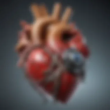
The four primary valves of the human heart—the mitral valve, tricuspid valve, pulmonary valve, and aortic valve—serve essential functions in the cardiovascular system. Each valve has a specific role in directing blood flow, ensuring the heart operates efficiently. Understanding these valves is critical for comprehending heart function overall. Their structure is finely tuned to withstand the rigors of constant blood circulation.
The significance of these valves cannot be understated. They prevent backflow, control pressure, and maintain adequate flow from one chamber to another. Any dysfunction can lead to severe complications, thus highlighting the importance of recognition and assessment of valve health.
Mitral Valve: Structure and Function
The mitral valve, located between the left atrium and left ventricle, has a unique structure. It consists of two leaflets attached to chordae tendineae that connect to papillary muscles. This design allows it to open and close effectively during the cardiac cycle. When the left atrium fills with blood, the mitral valve opens, allowing blood to flow into the left ventricle. Once the ventricle contracts, the valve closes, preventing blood from returning to the atrium.
The function of the mitral valve is vital. It ensures that oxygen-rich blood moves efficiently into systemic circulation. Any abnormalities, such as mitral valve prolapse or stenosis, can result in serious consequences, including heart failure.
Tricuspid Valve: Role in Circulation
The tricuspid valve sits between the right atrium and right ventricle. Composed of three cusps, this valve regulates blood flow from the atrium to the ventricle. During diastole, the tricuspid valve opens, allowing deoxygenated blood to enter the right ventricle from the right atrium. It subsequently closes during ventricular contraction to prevent backflow into the atrium.
Its role in circulation is linked to pulmonary circulation. The tricuspid valve ensures that blood reaches the lungs for oxygenation, making it critical for overall cardiovascular health. Disorders affecting this valve can lead to complications such as right-sided heart failure.
Pulmonary Valve: Mechanism of Action
The pulmonary valve is responsible for directing blood flow from the right ventricle into the pulmonary artery. It consists of three cusps and opens during ventricular contraction, allowing deoxygenated blood to flow to the lungs. The valve closes during the relaxation phase to prevent blood regurgitation into the ventricle.
This mechanism of action has significant implications for respiratory gas exchange. Proper function of the pulmonary valve is essential; issues such as pulmonary valve stenosis can impede blood flow, resulting in systemic complications and diminished oxygen supply.
Aortic Valve: Ensuring Unidirectional Flow
The aortic valve, positioned between the left ventricle and aorta, also contains three cusps. Its function is to maintain unidirectional flow of oxygenated blood to the body. Upon ventricular contraction, the aortic valve opens, permitting blood to exit into the aorta.
The closure of the aortic valve is equally important. It prevents backflow into the left ventricle during diastole. Dysfunction, such as aortic regurgitation, can lead to significant cardiovascular issues, affecting systemic circulation and overall heart efficiency.
Understanding these four valves and their functions provides essential insight into the workings of the heart. Each valve's structure and operation contribute intricately to the heart's overall performance, emphasizing their importance in maintaining health.
Physiological Function of Heart Valves
The physiological functions of heart valves are fundamental to the effective operation of the cardiovascular system. Understanding these functions sheds light on how the heart maintains optimal blood circulation and how various factors can influence this essential process. The heart valves prevent backflow, ensuring that blood moves smoothly through different chambers with each contraction. Their proper functioning is paramount to sustaining life and preventing complications associated with cardiovascular diseases.
Role in Blood Flow Regulation
Heart valves serve a critical role in regulating blood flow throughout the heart. Each valve can be likened to a gate that opens and closes based on pressure changes within the heart chambers. This regulation is vital after each heart contraction. When the heart muscle contracts, it creates pressure that forces blood through the valves. Each valve opens to allow blood to flow into the next chamber or blood vessel and then closes to prevent any backflow.
For example, the mitral valve opens to let oxygen-rich blood flow from the left atrium to the left ventricle. When the left ventricle contracts, the valve closes tightly to stop blood from flowing back into the left atrium. Similarly, the tricuspid valve facilitates blood flow from the right atrium to the right ventricle while preventing the backflow during contraction.
The careful choreography of opening and closing these valves is essential. Should any valve malfunction—such as narrowing (stenosis) or leakage (regurgitation)—the efficiency of blood flow can be compromised, leading to a variety of health issues.
"The precise function of heart valves is essential for maintaining the directional flow of blood, which is necessary for overall cardiovascular health."
Coordination with Heart Chambers


The heart operates as a synchronized unit. The coordination between the heart’s chambers and the valves is crucial for efficient blood circulation. Each heart chamber has a specific role, and the valves are the gateways ensuring that blood moves correctly at each phase of the cardiac cycle.
Firstly, during diastole, the heart chambers fill with blood. The atria receive blood from the body and lungs while the ventricles are relaxed. As the atria contract, blood is pushed into the ventricles through the open mitral and tricuspid valves. At this point, the aortic and pulmonary valves remain closed, preventing backflow from the arteries into the ventricles.
During systole, the ventricles contract, sending blood into the aorta and pulmonary artery. The aortic and pulmonary valves open to allow this flow, while the mitral and tricuspid valves close tightly, ensuring that blood does not return to the atria. This coordinated effort between heart chambers and valves is critical for effective pumping action, which is the essence of cardiac output—essential for life.
In summary, the physiological functions of heart valves are not only central to blood flow regulation but also vital for the coordination of heart chamber activities. Understanding these factors can lead to better insights into cardiovascular health and the implications of valve disorders.
Common Valvular Disorders
Common valvular disorders significantly affect cardiovascular health and can lead to severe complications. With the heart valves regulating blood flow, understanding these disorders is crucial for preventing adverse outcomes. They can impact anyone, regardless of age, making awareness of symptoms and treatment options vital. Factors such as genetic predisposition, lifestyle choices, and underlying health conditions can all contribute to the development of these disorders. Thus, any discussion about heart health must include common valvular disorders to provide a well-rounded perspective on cardiac function and risks.
Stenosis: Impeding Blood Flow
Stenosis occurs when a heart valve narrows, restricting blood flow. This condition can affect any of the four primary valves but is most common in the aortic and mitral valves. Consequently, the heart must work harder to pump blood through the narrow opening, leading to various complications. Symptoms of stenosis may include fatigue, shortness of breath, and chest pain, particularly during physical activity.
Causes of stenosis can range from congenital defects to age-related degeneration or rheumatic fever.
Diagnosis typically involves echocardiography, which helps visualize the valve structure and assess blood flow dynamics. In many cases, treatment may involve surgical interventions, such as valve replacement or repair. Understanding stenosis aids in early detection and management, thus preserving heart function.
Regurgitation: Backflow Issues
Regurgitation refers to the backflow of blood due to improper valve closure. This issue often occurs in the mitral and aortic valves, leading to increased pressure in the heart chambers. As a result, the heart becomes less efficient at pumping blood. Symptoms may include palpitations, fatigue, and systemic congestion, making it essential to recognize early signs.
Factors contributing to regurgitation include damage from previous heart conditions, age, or congenital issues. Diagnosis is usually confirmed through echocardiography, revealing changes in valve structure. Treatments may include medication to manage symptoms or surgery when the condition is severe. Addressing regurgitation is critical for maintaining healthy circulation.
Infective Endocarditis
Infective endocarditis is a serious infection of the heart valves or inner lining. It is often due to bacteria entering the bloodstream and settling on damaged valves. This disorder can result in life-threatening complications such as heart failure or embolic events, emphasizing the need for accurate diagnosis and prompt treatment.
Symptoms can be non-specific, such as fever, chills, or fatigue, which may delay diagnosis. Risk factors include pre-existing valvular heart disease, intravenous drug use, and recent medical procedures. Diagnosis involves blood cultures and echocardiography to identify the infective organism and assess valve damage.
Treatment typically requires prolonged antibiotic therapy, and in severe cases, surgery may be necessary to remove infected tissue. Understanding this disorder helps reinforce the importance of cardiovascular hygiene, especially for those at risk.
Understanding common valvular disorders, such as stenosis, regurgitation, and infective endocarditis, is essential for effective cardiovascular management and prevention strategies.
By integrating knowledge about common valvular disorders, readers can foster a better awareness of their cardiovascular health.
Diagnosis of Valvular Disorders
Diagnosing valvular disorders is critical in understanding heart health and function. The heart valves play an essential role in maintaining proper blood flow, and any dysfunction can lead to serious health complications. Accurate diagnosis allows for timely interventions, which can range from monitoring the condition to implementing life-saving treatments. The process involves a combination of patient evaluation, symptom assessment, and advanced imaging techniques. It is essential for healthcare providers to recognize the signs and symptoms of valvular issues.
Echocardiography
Echocardiography is an invaluable tool in the diagnosis of heart valve disorders. This non-invasive imaging technique uses sound waves to create moving images of the heart. It provides detailed visualization of valve structure and function. During an echocardiogram, healthcare professionals can observe how blood flows through the heart valves during different phases of the cardiac cycle.
Some of the key benefits of echocardiography include:
- Real-time imaging: This allows immediate assessment of blood flow and valve function.
- Evaluation of heart chambers: Echocardiography can also assess chamber size and function to provide a holistic view of cardiac health.
- Detection of abnormalities: Any structural or functional abnormalities such as stenosis or regurgitation can be identified.
In summary, echocardiography plays a crucial role in making informed decisions regarding the management of valvular disorders.
Cardiac MRI
Cardiac MRI is another important diagnostic tool for assessing heart valve disorders. Unlike echocardiography, cardiac MRI uses magnetic fields and radio waves to produce detailed images of the heart. This imaging modality excels in evaluating complex heart conditions. It provides clear illustrations of the heart's anatomy, including valves.
The advantages of cardiac MRI include:


- High-resolution images: MRI offers superior detail, making it easier to identify subtle defects not visible on echocardiograms.
- Functional assessment: This technique can evaluate how well the heart is functioning by measuring blood flow and the contractility of the heart muscle.
- Comprehensive data: The ability to assess both structure and function limits the need for invasive procedures.
Ultimately, cardiac MRI complements other diagnostic methods and enhances the understanding of valvular abnormalities.
The combination of echocardiography and cardiac MRI provides a robust framework for diagnosing valvular disorders, enabling clinicians to devise appropriate treatment plans.
Accurate diagnosis is foundational in the treatment of heart valve disorders, ensuring patients receive optimal care tailored to their specific needs.
Treatment Approaches for Valve Issues
In the realm of cardiovascular health, understanding treatment approaches for valve issues is paramount. The human heart's valves are intricate structures that require proper management when disorders arise. Effective treatment not only alleviates symptoms but also restores normal heart function. Here, we explore various methods employed in addressing valve problems, ensuring a comprehensive overview for those interested in this critical aspect of cardiology.
Medical Management
Medical management of valve disorders often serves as the first line of treatment. This approach may include medications to manage symptoms and prevent disease progression. The following are commonly prescribed:
- Anticoagulants: These drugs help reduce the risk of blood clots, particularly in patients with valve-related conditions like atrial fibrillation.
- Beta-blockers: Often used to treat high blood pressure and heart rhythm issues, beta-blockers can ease the heart's workload and improve overall heart function.
- Diuretics: These medications help reduce fluid retention, a common issue in valvular disease, improving symptoms such as swelling and shortness of breath.
While medical management may effectively control symptoms, it does not resolve structural issues with the valves. Therefore, it is crucial to evaluate patients for potential surgical interventions if symptoms persist or worsen.
Surgical Interventions
When valve disorders reach a critical stage, surgical interventions become necessary. Surgery aims to repair or replace malfunctioning valves, restoring proper blood flow. Two primary surgical approaches exist:
- Valve Repair: This procedure aims to fix the existing valve, which is often preferable to replacement. Surgical techniques vary depending on the specific valve and nature of the defect. Repair can include tightening or reshaping the valve to improve function.
- Valve Replacement: If a valve is severely damaged, replacement becomes essential. Surgeons may use biological valves, made from animal tissue, or mechanical valves, which are man-made. Each option has distinct benefits and risks, requiring careful consideration based on the patient's age, health status, and lifestyle.
"Early surgical intervention can significantly improve outcomes for patients with severe valvular disease."
Both surgical options require thorough preoperative evaluations and postoperative care to ensure successful recovery and function.
Innovations in Valve Replacement
The field of valve replacement has witnessed remarkable advancements, focusing on improving patient outcomes and minimizing recovery times. One such innovation is the transcatheter aortic valve replacement (TAVR), a less invasive method compared to traditional surgical options. This technique utilizes a catheter to insert a new valve through a blood vessel, allowing treatment without opening the chest.
- Benefits of TAVR:
- Reduced recovery time
- Lower risk of complications
- Applicable for patients considered high-risk for open-heart surgery
Another promising development is the creation of 3D-printed heart valves, tailored specifically to individual patient anatomies. This technology has the potential to enhance the compatibility and functionality of replacements, leading to better long-term outcomes.
As researchers continue to explore cutting-edge solutions, the future of valve treatment looks promising. Ongoing clinical trials and studies are essential for understanding the full capabilities and limitations of these innovations.
In summary, treatment approaches for valve issues comprise a combination of medical management, surgical interventions, and innovative replacements. Each method has its role in optimizing cardiac health and ensuring the heart's valves function effectively.
Closure: The Significance of Understanding Heart Valves
A detailed comprehension of the heart valves is critical for both health professionals and patients alike. These structures play a pivotal role in maintaining unidirectional blood flow, ensuring proper systemic circulation. Understanding the anatomy and function of each valve aids in the identification of potential issues that can arise within the heart. Heart valve diseases can severely impact a patient's quality of life, making it essential for anyone involved in cardiovascular care to be well-versed in the mechanisms and purposes of these valves.
The knowledge of how valves operate and their connection with heart chambers contributes to better diagnosis and treatment options for patients. For instance, recognizing the signs and symptoms of common valvular disorders such as stenosis or regurgitation allows for timely medical intervention. Moreover, understanding advancements in treatments, from medical management to surgical options, empowers professionals and patients to engage in informed discussions about care strategies.
"Health care professionals and patients must work together to advance understanding and treatment of heart valve diseases."
Additionally, education on heart valve function encourages timely monitoring and maintenance of cardiovascular health. As research continues to evolve, the integration of new technologies in diagnostic tools and surgical techniques will become increasingly critical. Thus, the significance of understanding heart valves extends beyond just academic knowledge; it is vital for clinical application and improved patient outcomes.
Summary of Key Points
- Heart valves are essential for ensuring proper blood circulation.
- Each valve serves a unique function in the cardiac cycle.
- Disorders can lead to serious health complications.
- Timely diagnosis and informed treatment options are crucial.
- Continued research and education are essential for improved health outcomes.
Call for Continued Research
The field of cardiovascular health is ever-evolving, and continued research into heart valves is imperative. As new knowledge emerges about the complexity of heart valve function and associated disorders, it paves the way for innovative treatments and technologies. Studies focusing on the molecular mechanisms of valve diseases can lead to breakthroughs in prevention and management strategies. Furthermore, understanding patient-specific factors that influence valve disease progression remains a priority.
Engaging with multidisciplinary teams can enhance research efforts. Collaboration among cardiologists, surgeons, and biomedical engineers can foster advancements in valve repair or replacement techniques. Additionally, exploring patient education initiatives can improve awareness about valve issues and signify the importance of early symptoms recognition.
In summary, ongoing research is essential to advance our understanding of heart valves, their disorders, and treatments. Only through continued inquiry and innovation can we ensure the best possible outcomes for patients.


